Επιλεγμένες Επιστημονικές Δημοσιεύσεις

Penetrating internal jugular vein injury with massive bleeding

S. Papadoulas et al. Pol J Thoracic and Cardiovascular Surg 2025; 22 (2): 126-128. doi: https://doi.org/10.5114/kitp.2025.152193. Department of Vascular Surgery, University of Patras Medical School, Patras, Greece
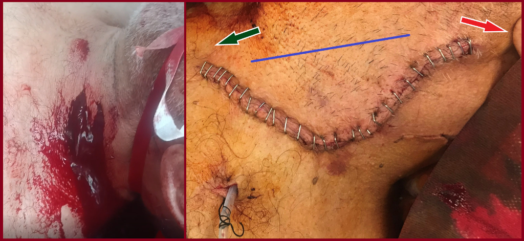
The neck is a complex anatomical area that occupies unique vital structures. They can be severely damaged in any perforating neck trauma, leading to considerable morbidity and mortality. Non-iatrogenic internal jugular vein (IJV) injuries are rare, and no specific guidelines exist regarding their optimal treatment. We present a rare case of an extended IJV transaction and massive exsanguinations through a perforating neck trauma.

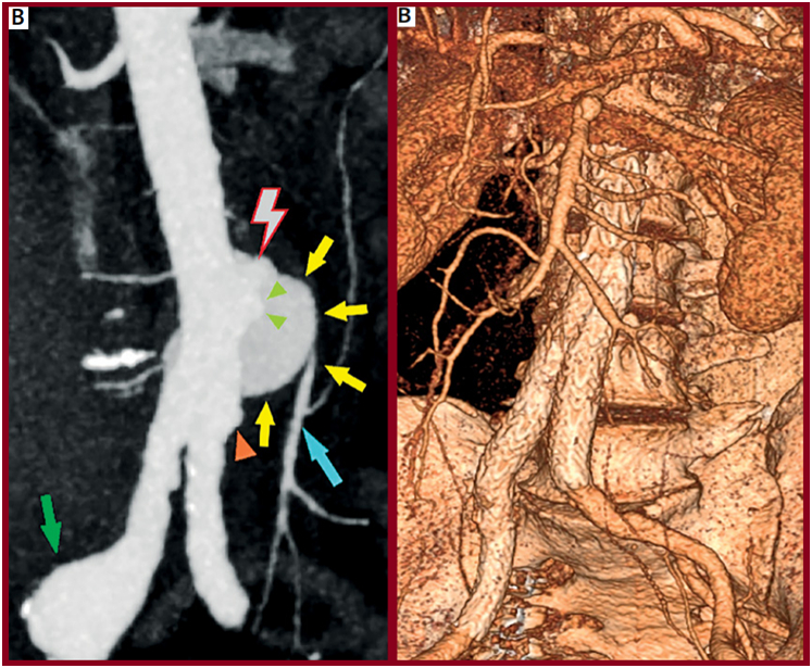
Spontaneous avulsion of the inferior mesenteric artery in a neurofibromatosis type 1 patient: a case-based review

S.Papadoulas et al Arch Med Sci Atheroscler Dis 2025; 10: e48–e68 doi:https://doi.org/10.5114/amsad/205022. Department of Vascular Surgery, University of Patras Medical School, Patras, Greece

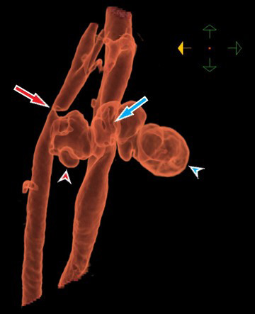
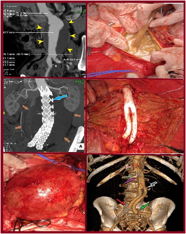
We present the case of successful endovascular abdominal aortic aneurysm repair (EVAR) in a 55-year-old male who presented with a ruptured infrarenal aortic pseudoaneurysm, formed after spontaneous avulsion of the inferior mesenteric artery. The avulsion occurred after lifting a heavy object. Although aortic endografting is not the first option in patients with hereditary disorders due to aortic friability and concerns about long-term durability, it is valuable in urgent cases due to lower morbidity and mortality, even as a bridging procedure. This policy is further supported by the fact that the alternative open reconstruction has been invariably associated with hemostasis issues and poor outcomes due to arterial fragility and inability to construct safe anastomoses. Finally, we present a current literature review regarding abdominal aortic and iliac pathology in patients with neurofibromatosis type 1 focusing on the type of the vascular lesion, method of repair and outcome.
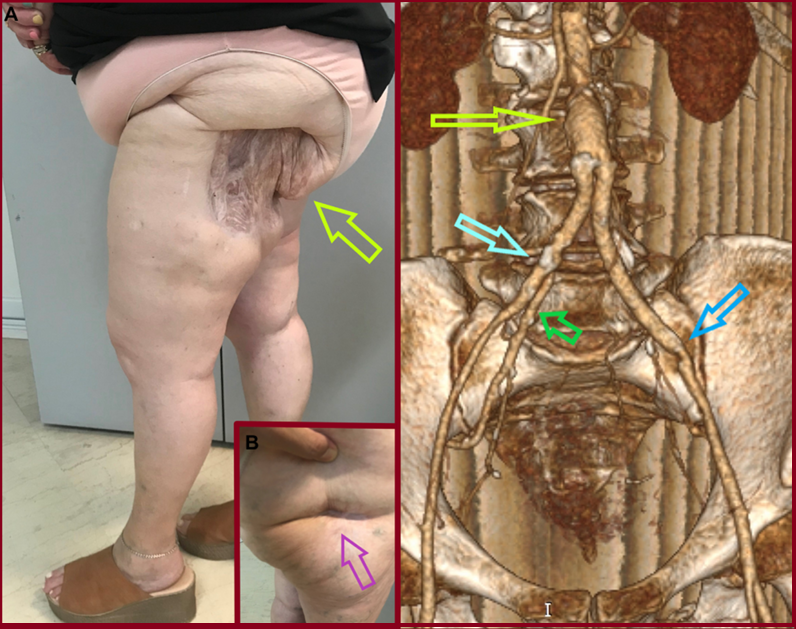
Gluteal ischemic gangrene due to chronic aortoiliac occlusive disease

C. Pitros, S. Papadoulas et al. J Vasc Surg Cases Innov Tech 2025;11:101792. Department of Vascular Surgery, University of Patras Medical School, Patras, Greece

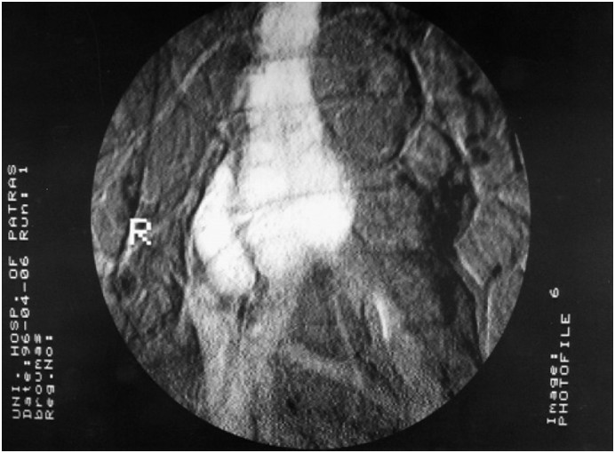
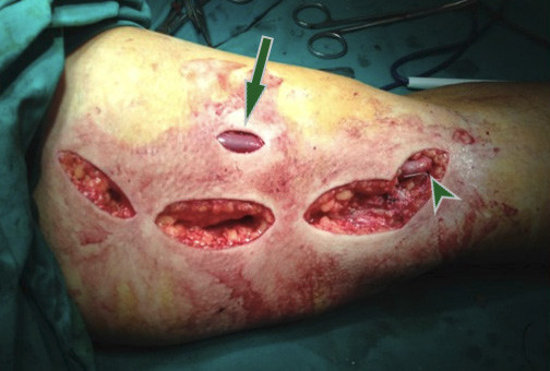
Gluteal gangrene rarely complicates interventional procedures such as angiographic embolization in the pelvis or may develop postoperatively after open ligation or endovascular covering or embolization of one or mainly both internal iliac arteries in abdominal aortic surgery if collateral circulation is compromised. On the other hand, gluteal gangrene as a primary manifestation of chronic aortoiliac occlusive disease is very exceptional in literature. We present a patient with atherosclerotic aortoiliac obstruction and a necrotic eschar on her left buttock treated with aortobiiliac bypass after digital subtraction angiography. Internal iliac artery revascularization, even contralaterally, is crucial for the healing of necrotic tissue in these patients.
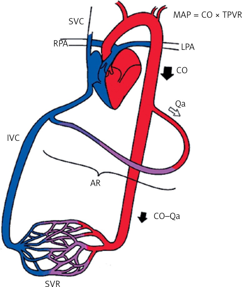
Cardiac complications of arteriovenous access: a narrative review from a multidisciplinary team perspective

Stathopoulou, S. Papadoulas et al. Arch Med Sci Atheroscler Dis 2024; 9: e217–e225. Department of Vascular Surgery, University of Patras Medical School, Patras, Greece

Although cardiovascular disease is common among hemodialysis patients, arteriovenous access creation has been invariably implicated in the evolution of adverse cardiac outcomes or deterioration of pre-existing cardiovascular disease. In most cases, these effects are subclinical but with potential underlying echocardiographic findings. Compared with grafts, arteriovenous fistulas are implicated more often, due to the progressively increased flow from the continuous dilatation of the venous outflow tract in the long term. The increasing flow is in the majority of patients well tolerated by cardiac adaptive alterations. However, the clinical impact is based on the balance between the amount of flow volume and the patient’s cardiac reserves. Having extensively reviewed the existing English literature, we present the pathophysiology and the different types of cardiovascular complications, the indications, types, and efficacy of flow-restrictive procedures in the con text of a high-flow AVF, as well as some precautions and considerations for AVF creation in high-risk patients.
Penetrating carotid artery injury

V. Argitis, S. Papadoulas et al. Hellenic Journal of Vascular and Endovascular Surgery | Volume 6 – Issue 2 – 2024. Department of Vascular Surgery, University of Patras Medical School, Patras, Greece

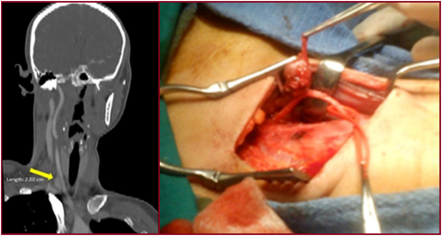
A 27-year-old male presented with a left cervical pulsative mass after penetrating trauma with a knife. No external bleeding was observed despite the presence of an open skin incision over the sternocleidomastoid muscle. Urgent Computed Tomography Angiography (CTA) revealed a partial transverse transection of the left common carotid artery (CCA) and the adjacent internal jugular vein (IJV) surrounded with a cervical hematoma due to gross extravasation from both vessels (Figure). In operating theatre, after a standard open approach for carotid endarterectomy, the CCA and IJV were clamped. The CCA was totally transected at the injury site, trimmed and re-anastomosed in an end-to-end fashion. The IJV was primarily sutured, and the hematoma was evacuated. The postoperative care was uneventful, and the patient was discharged on the 4th postoperative day. She was prescribed single antiplatelet and antibiotic treatment
Late post-EVAR abdominal aortic aneurysm rupture: a meta-analysis study
Papadoulas SI et al. Arch Med Sci Atheroscler Dis. 2024;9:e152–e164. Department of Vascular Surgery, University of Patras Medical School, Patras, Greece
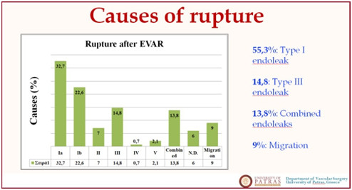
This study presents updated information on post-endovascular aneurysm repair (EVAR) late aortic rupture (LAR) as the data in the literature are limited. It comprises a meta-analysis based on the recent evidence regarding the incidence, causes, treatment outcomes, and prognosis of post-EVAR. A meta-analysis was conducted according to the PRISMA (Preferred Reporting Items for Systematic Reviews and Meta-Analyses) guidelines. Studies were identified by searching electronic databases and scanning bibliographic references from 1991 to April 1, 2023. Our analysis provided evidence that the most common causes of rupture after EVAR were type Ia and Ib Endoleaks (Els). Post-rupture mortality after EVAR was high (35.6%) and comparable to the morbidity of de novo ruptures. Endovascular repair appears to have better results compared to conversion to open repair. A significant number of patients had prior endovascular reoperations and inadequate follow-up. Patient compliance with the surveillance protocol is mandatory

The perivascular hypodense rim in carotid body tumor surgery
Mila E, Papadoulas SI et al. Hellenic Journal of Vascular and Endovascular Surgery 2024;Volume 6:Issue 1. Department of Vascular Surgery, University of Patras Medical School, Patras, Greece
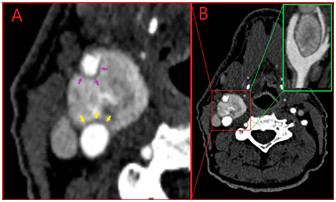
A 57-year-old male, current smoker presented with a threeyear history of a right neck mass progressively enlarging. CTA established the diagnosis of a Shamblin II carotid body tumor (CBT) 2.5×3.0cm in size. An inactive pheochromocytoma was also depicted in the left adrenal gland, 1cm in size. The internal carotid artery (ICA) was partially engaged in the tumor at 140o along its circumference while the external carotid artery (ECA) at 245o. A perivascular hypodense rim was apparent (Fig 1). The patient underwent surgical excision of the CBT with partial clamping of the ECA to aid haemostasis (Fig 2). Postoperative course was uneventful, and the patient was discharged on the 3rd postoperative day. The presence of a perivascular hypodense rim in our patient indicates safe tumor resectability without need for ICA reconstruction or ECA excision. It corresponds to the fat plane normally lying around the arteries. Loss of this avascular plane indicates infiltration of the vessel wall by the tumor, making subadventitial dissection impossible. In a recent series from Jasper et al, the absence of this rim displayed as loss of tumor adventitia interface was significantly different between the Shamblin groups (33.3% in Shamblin I, 60% in II and 95,2% in III)1 . Consequently, lack of this radiological sign increases the likelihood for tumor adherence to ICA needing arterial reconstruction and higher morbidity is anticipated (e.g., strokes and cranial nerve palsies).

The ‘hook-sign’ in the Median Arcuate Ligament Syndrome

Papadoulas SI, et al. Hellenic Journal of Vascular and Endovascular Surgery 2020;Volume 2:Issue 3. Department of Vascular Surgery, University of Patras Medical School, Patras, Greece

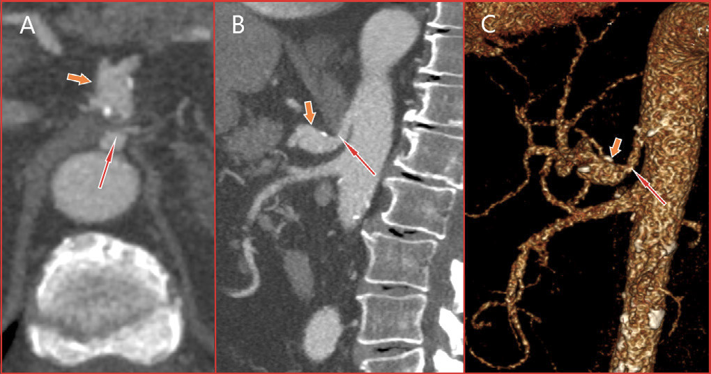
An 83-year-old male, former smoker with a medical history of hypertension and a thoracic aortic aneurysm was presented complaining of itching and edema of his legs. Symptoms were attributed to tactile skin purpura. An abdominal computed tomography (CT) scan was performed to rule out a para-neoplasmatic syndrome. An asymptomatic compression of the celiac artery by the median arcuate ligament (MAL) along with celiac artery kinking and a significant post-compression dilatation (15x15mm in size) was depicted. A following CT An-giogram (CTA) delineated the arterial anatomy and suggested the co-existence of an asymptomatic median arcuate ligament syndrome (MALS), (Figure 1). Median arcuate ligament syndrome was first described in 1917 by Lipshutz who noticed the overlapping of the artery by the diaphragmatic crura in an autopsy. It is a rare condition characterized by recurrent episodes of epigastric pain, nausea, diarrhea, and weight loss. The pain tends to be postprandial with a recession when leaning forward. Symptomatology is attributed to celiac artery compression by the arch-shaped ligament. Lower origin of the celiac artery or superior position of the MAL may predispose to MALS. The distortion of the CA centerline creates the ‘hook-sign’ (Figure 2). Sometimes, a neuropathic pain may be apparent due to ischemia or compression of the celiac ganglion. Current treatment consists of laparoscopic ligament transection followed by celiac artery angioplasty. A post-stenotic aneurysm larger than 2cm is generally
repaired by covered-stents or open surgery.
Multiple skip incisions technique for two-staged basilic vein transposition: a good alternative to the standard single long incision

Papadoulas SI, et al. Hellenic Journal of Vascular and Endovascular Surgery 2020;Volume 2:Issue 3. Department of Vascular Surgery, University of Patras Medical School, Patras, Greece

A 58-year-old-male on hemodialysis underwent a two-staged tunneled basilic vein transposition with three skip incisions. Initially, preoperative color-duplex mapping was performed and a standard brachio-basilic arteriovenous fistula with the median cubital vein was accomplished. Six weeks later, after the arterialization of the basilic vein, the second stage took place after skin marking of basilic vein’s location by ultrasound. Under local anesthesia, the basilic vein and the cubital vein were dissected free from the arterial anastomosis up to the confluence with the axillary vein, using hook/right-angle retractors to dissect the vein under the skin. The vein was transected peripherally, tunneled subcutaneously in a new lateral route, after a small skin incision in the mid-upper arm, and re-anastomosed. The incisions were closed in a standard fashion. The access is still functional during the last six years. Multiple skip incisions compared to a single long incision have a better cosmetic result and cause less tissue damage. A single incision offers easier vein dissection, needs less operative time but leaves back a longer scar. Multiple skip incisions technique would have the theoretical advantage of reduced postoperative pain, oedema and surgical site infection/dehiscence rates. This is supported in some reports1 but in others, although these complications were encountered infrequently, the difference did not reach statistical significance.
Recurrence of a Resected Carotid Body Tumor Presenting as a Vagal paraganglioma

Papadoulas SI, et al. Hellenic Journal of Vascular and Endovascular Surgery 2020;Volume 2:Issue 3. Department of Vascular Surgery, University of Patras Medical School, Patras, Greece

Purpose: To present a rare case of carotid body tumor recurrence presenting as a vagal paraganglioma. Vagal paragangliomas represent. Case report: An asymptomatic 25-year-old female was diagnosed with a vagal paraganglioma 3 years after an ipsilateral carotid body tumor resection. which had been fully excised as was shown by the pathology specimen examination and a recent Computed Tomography Angiography (CTA). Preoperative presumed diagnosis was based only on duplex scan and CTA. No biopsy was performed because it is contraindicated due to high vascularity of these tumors. Vagal Paraganglioma resection was easily performed because the patient was thin with a clean surgical field locating just distal to the previews operated area. Pathology specimen was conclusive for paraganglioma and his conjunction with the vagus nerve found on operation was indicative for a vagal paraganglioma. The patient remains without recurrence during the last 4 years. Conclusion: This case emphasizes the necessity of a strict postoperative follow-up protocol after resection of paragangliomas, as recurrence is always possible.
Endotension as a Rare Complication to Endovascular Abdominal Aortic Aneurysm Repair.

Papadoulas SI, et al. Innovations (Phila) 2023;18(5):498-502. doi: 10.1177/15569845231191129. Epub 2023 Aug 8. PMID: 37551670. Department of Vascular Surgery, University of Patras Medical School, Patras, Greece
Endotension remains an enigmatic rare cause of endovascular abdominal aortic aneurysm repair failure leading to aneurysm growth and/or rupture. We present a patient with a long-standing endotension treated with open reconstruction and graft explantation. We also provide a unique clinical video, which gives a key view of the intrasac operative findings.

Myasthenia Gravis and Abdominal Aortic Aneurysm: A Rare Combination

Papadoulas SI, et al. Aorta (Stamford). 2023 Mar 20. doi: 10.1055/a-2051-7678. PMID: 36940930. Department of Vascular Surgery, University of Patras Medical School, Patras, Greece

Abdominal aortic aneurysm in a patient with myasthenia gravis (MG) is extremely rare. We present a 64-year-old male with MG and an asymptomatic abdominal aortic aneurysm treated endovascularly. After extubation, he suffered a cardiac arrest due to an acute myocardial infarction. Cardiopulmonary resuscitation and a primary coronary angioplasty led to a satisfactory outcome. Special care is needed due to higher rates of postoperative complications in these patients.
Endovascular Repair of an Inflammatory Abdominal Aortic Aneurysm Combined with a Congenital Pelvic Kidney: Case Report and Literature Review.
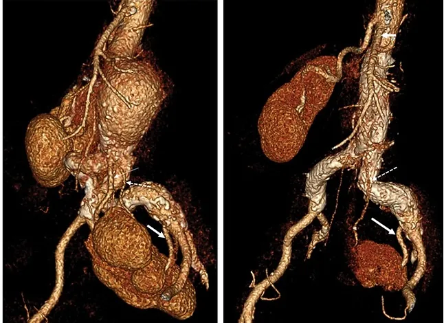
Papadoulas S, et al. Aorta (Stamford). 2022 Jun;10(3):135-140. doi: 10.1055/s-0042-1748961. Epub 2022 Nov 1. PMID: 36318935; PMCID: PMC9626033. Department of Vascular Surgery, University of Patras Medical School, Patras, Greece
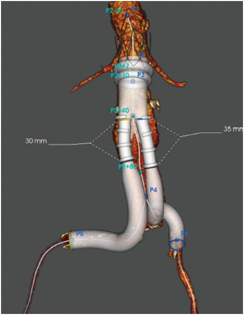
The coexistence of an abdominal aortic aneurysm and a congenital pelvic kidney is extremely rare. We present a 66-year-old male with an inflammatory aneurysm and an aberrant origin of the superior mesenteric artery. The inflammatory infrarenal abdominal aortic aneurysm with a congenital left pelvic kidney was successfully treated with endovascular repair. Coverage of one out of the two renal ectopic arteries was performed, without clinical evidence of renal function impairment.

Superficial temporal artery pseudoaneurysm after blunt trauma: A case series
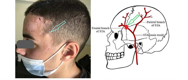
Christos Pitros, Spyros I. Papadoulas et al. Hellenic Journal of Vascular and Endovascular Surgery, Volume 4, Issue 3, 2022. Department of Vascular Surgery, University of Patras Medical School, Patras, Greece
Purpose: To describe our experience with the presentation and management of traumatic superficial temporal artery pseudoaneurysms in our institution. Small case series description: Reviewing all patient’s records during the last 20 years we identified 3 cases with a superficial temporal artery pseudoaneurysm. Two patients had suffered a motorcycle accident and presented with blunt and/or penetrating skin injuries on the head remote of the site of the aneurysm. No helmet was used. The remaining patient presented with skin necrosis on the aneurysm indicating a direct blunt arterial injury after a minor fall. No other severe injuries were apparent. After a diagnostic colour duplex they underwent aneurysm excision and ligation of the superficial temporal artery under local anesthesia. Conclusion: In our patients’ temporal artery pseudoaneurysms were developed after direct arterial transection or traction due to a nearby head injury. Diagnosis was based on clinical examination and confirmed by ultrasound. Treatment consisted of aneurysm excision.

A case report of surgical repair of a post-catheterization radial pseudoaneurysm.
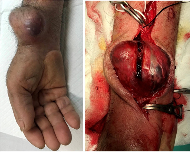
Papadoulas SI, et al. Pan Afr Med J. 2022 Mar 31;41:261. doi: 10.11604/pamj.2022.41.261.29725. PMID: 35734330; PMCID: PMC9187997. Department of Vascular Surgery, University of Patras Medical School, Patras, Greece
We report an 83-year-old patient with a huge post-catheterization right radial pseudoaneurysm, presented 17 months after a coronary angiography. Cases of radial post-catheterization pseudoaneurysms with a similar size are scarce in the literature. Delay in presentation led to painful skin ischemia due to tension, a sign of imminent rupture, which is also rare in the literature. Symptomatology included severe wrist pain and clinical signs consisted of a pulsatile painful mass in the right distal forearm. Management consisted of surgical excision and ligation of the radial artery in an urgent base. This case emphasizes the need for early diagnosis and management of post-catheterization pseudoaneurysms as delay may lead to severe enlargement with skin necrosis and imminent rupture. Ligation of the radial artery is an acceptable option when reconstruction of the artery is troublesome, provided that the palmar arch remains patent.

Papadoulas SI, et al. Treatment options for dialysis access steal syndrome.
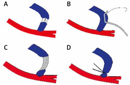
Kardiochir Torakochirurgia Pol. 2022 Sep;19(3):141-145. doi: 10.5114/kitp.2022.119762. Epub 2022 Oct 8. PMID: 36268490; PMCID: PMC9574582. Department of Vascular Surgery, University of Patras Medical School, Patras, Greece
Vascular access-induced limb ischemia is a known complication of arteriovenous fistulas and grafts. Many techniques have been adopted to prevent steal in high-risk patients and to treat steal in cases of moderate ischemia not controlled with conservative management. A major factor guiding treatment is access flow volume. Management is different when ischemia is combined with the excessive flow in contrast to the combination with normal flow. We describe the most popular techniques encountered in the English literature as a part of a stepwise approach to treating dialysis access steal syndrome. In absence of ischemia, when cardiac issues emerge due to extreme access flow volumes, some of these techniques are also used to decrease flow and protect the heart. Patient’s history, focused clinical examination, color duplex ultrasound examination, pulse oximetry and an angiogram are essential tools to approach this entity.

Patient Transfer with Kocher Forceps on the Axillary Artery: A Rare Case of Ongoing Iatrogenic Vascular Injury.
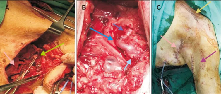
Seretis C, Spyros Papadoulas et al. Vasc Specialist Int. 2022 Mar 31;38:10. doi: 10.5758/vsi.220010. Department of Vascular Surgery, University of Patras Medical School, Patras, Greece
Iatrogenic trauma of the axillary artery by non-vascular surgeons can occur during various general surgical procedures such as resection of soft tissue tumors or axillary lymph node clearance. Prompt recognition, appropriate initial management, and rapid transfer to a tertiary vascular surgery service, if needed, are key steps to ensuring patient safety. Here we present a case of iatrogenic axillary artery injury during the resection of a recurrent soft tissue tumor in a local hospital. The desperate application of a Kocher clamp on the bleeding axillary artery by the operating general surgeons controlled the bleeding but led to further arterial damage. The patient was transferred to our tertiary hospital, where the arterial injury was repaired using a vein interposition graft. Apart from the encountered intraoperative technical challenges, this case highlights the need for broader training of nonvascular specialist surgeons on the core principles of basic vascular surgical techniques and oncovascular surgery.

Treatment of Dialysis Access Steal Syndrome with Concomitant Vascular Access Aneurysms
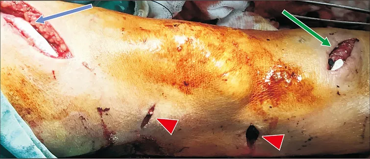
Spyros Papadoulas et al. Vasc Specialist Int. 2022 Mar 31;38:11. doi: 10.5758/vsi.220006. Department of Vascular Surgery, University of Patras Medical School, Patras, Greece
Limb ischemia is a known complication of vascular access that may appear early postoperatively or after years. Over the last few decades, various techniques based on different physiological mechanisms have been used for treatment. A standardized treatment does not exist, and must be individualized based on the flow volume, and the type and location of the access. True and false vascular access aneurysms are another common complication of arteriovenous fistulas, which develop because of venous hypertension or repeated needling. Evidence in the literature regarding treatment of patients with steal syndrome and concomitant true arteriovenous aneurysms is scarce. A female with a brachiocephalic fistula complicated by steal syndrome and vascular access aneurysms was treated successfully with tapered graft placement and aneurysm exclusion.

Short interposition grafting for dialysis-access steal syndrome treatment.

Papadoulas S et al. BMJ Case Rep. 2022 Feb 28;15(2):e248446. doi: 10.1136/bcr-2021-248446. Department of Vascular Surgery, University of Patras Medical School, Patras, Greece
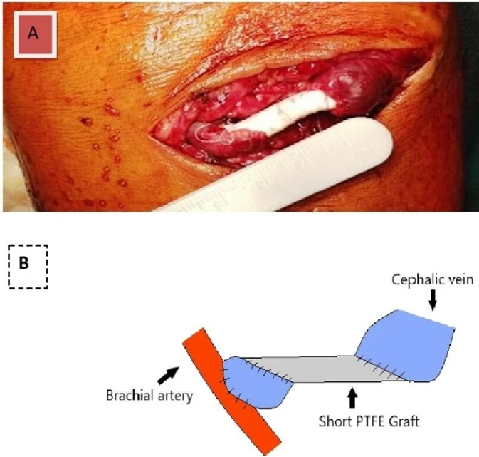
A 60-year-old man on chronic haemodialysis presented with access-related severe ischemia of the hand 4 years after the creation of a left brachiocephalic arteriovenous fistula. The fingers were painful, he was pale, and skin ulceration was evident on the thumb. Concomitant diseases included arterial hypertension and diabetes. Doppler signals were severely attenuated in the forearm arteries but returned to normal after digital compression of the fistula. He underwent colour duplex examination, which revealed brachial artery flow of 2600 mL/min. The anastomosis was 8 mm wide, and the diameter of the proximal cephalic vein was approximately 1.5 cm. A diagnosis of access-related steal syndrome (grade 4a) due to hyperfunctioning brachiocephalic fistula was made. Digital subtraction angiography (DSA) ruled out the presence of arterial stenoses proximal to the fistula that could affect inflow. In addition, no stenoses were detected distally that could increase peripheral resistance. We performed ligation of a major cephalic side branch to restrict overflow from the fistula, but without apparent benefit. We then subjected the patient to a more invasive procedure to restrict flow. Under local anaesthesia, we inserted a short expanded polytetrafluoroethylene (ePTFE) graft (Gore Intering) with a diameter of 6 mm and a length of 3 cm extending 1 cm beyond the anastomosis (figure 1A,B). Because of the discrepancy between the diameters, the anastomoses were created obliquely and performed, so that the resulting angles in the anastomoses corresponded to the upward rotation of the cephalic vein. Postoperatively, brachial flow decreased to 1000 mL/min, the patient’s symptoms disappeared and the ulcer eventually healed. Three weeks later, the patient began haemodialysis through the fistula. For the next 7 years, the fistula was used for haemodialysis without recurrence of steal until the patient died of cancer….

Iatrogenic tibial arteriovenous fistula after Fogarty balloon catheter graft thrombectomy

Spyros Papadoulas et al. Clin Case Rep. 2021 Nov 9;9(11):e05050. doi: 10.1002/ccr3.5050. Department of Vascular Surgery, University of Patras Medical School, Patras, Greece
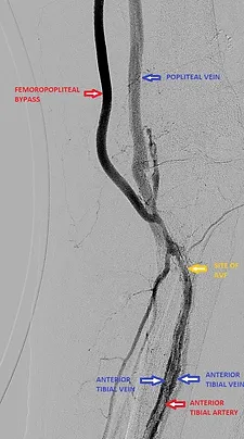
A 75‐year‐old male presented with an immediately threatened grade IIb acute ischemia of the left leg due to thrombosis of a femoro‐infrapopliteal prosthetic bypass graft. After an urgent Computed Tomography Angiography, an urgent graft thrombectomy was performed using a 5 Fr Fogarty catheter, which had a troublesome distal passage, causing a tibial A‐V fistula.

Custom-Made Bifurcated Prosthetic Graft for Aortoiliac Aneurysm Repair
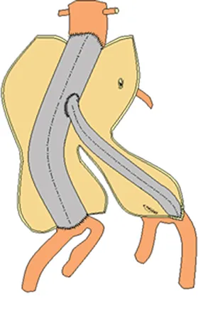
Spyros Papadoulas et al. Aorta (Stamford) 2021 Apr;9(2):88-91. doi: 10.1055/s-0041-1725090. Department of Vascular Surgery, University of Patras Medical School, Patras, Greece
Revascularization of the internal iliac artery during open repair of aortoiliac aneurysms can be challenging, especially if there is a significant distance between the orifices of the internal and external iliac arteries owing to common iliac aneurysmal dilatation. We describe a technique involving insertion of an 18-mm tube graft between the proximal aortic neck and aneurysmal common iliac artery bifurcation. Revascularization of the contralateral external iliac artery is accomplished through an 8-mm side arm graft.

Free-Floating Thrombus in the Distal Internal Carotid Artery Causing a Stroke
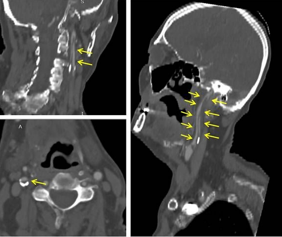
Spyros Papadoulas et al. Int J Angiol. 2021 Jun;30(2):170-172. doi: 10.1055/s-0040-1720973. Department of Vascular Surgery, University of Patras Medical School, Patras, Greece
We present a patient suffering from a stroke with a free-floating thrombus extending up to the distal internal carotid artery. The thrombus was totally resolved after a 2-week anticoagulation regimen without leaving behind any severe residual stenosis in the carotid bulb. The optimal treatment of this rare condition remains uncertain. We report some important treatment strategies that have been used in the literature, emphasizing the anticoagulation as the mainstay of therapy. Immediate surgical and interventional manipulations carry the risk of thrombus dislodgement and embolization and should be considered if there are recurrent symptoms despite medical management.

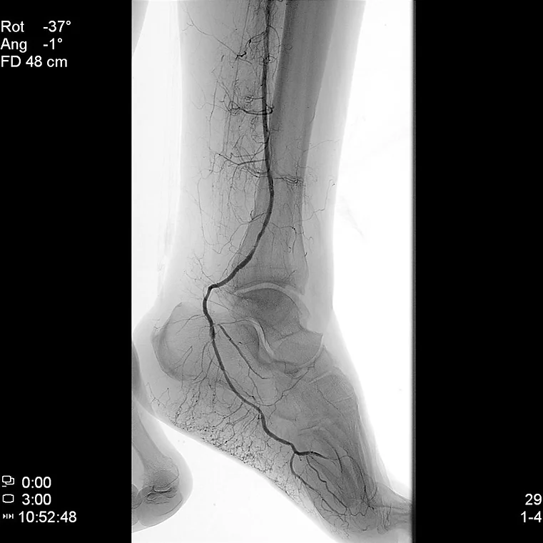
Adjunctive vacuum-assisted aspiration thrombectomy in a patient with acute limb ischaemia and peronea arteria magna
Spyros Papadoulas et al. BMJ Case Rep 2021 Aug 17;14(8):e245490. doi: 10.1136/bcr-2021-245490. Department of Vascular Surgery, University of Patras Medical School, Patras, Greece
A 63-year-old woman presented with acute left foot ischaemia with pain, sensory loss and moderate motor deficit. She was a heavy smoker with arterial hypertension, hyperlipidaemia and a history of left breast cancer 15 years ago. She urgently underwent a standard Fogarty embolectomy through a left groin common femoral artery incision under local anaesthesia. An arterial embolus with minimal amount of fresh thrombus was retrieved. The leg regained partial mobility and sensation, but the forefoot remained cold, pale and painful. Urgent intraoperative digital subtraction angiogram (DSA) is not normally performed in our department due to staff and equipment problems. The patient underwent a DSA in the Interventional Radiology Suite postoperatively where the equipment and experience are highly available. It revealed a dominant peroneal artery that was occluded above the level of malleolus with a completely deserted foot. Beyond this level, no vessel was opacified (figure 1A). Anterior tibial artery was hypoplastic but patent until mid-calf. An image-guided percutaneous vacuum-assisted aspiration thrombectomy with the INDIGO/PENUMBRA catheter was performed (figure 1B). Through the peroneal artery, using a 6F catheter, thrombus was retrieved from the plantar vessels down to the midsole, restoring normal vessel patency (figures 2 and 3). Posterior tibial pulses were restored, the foot immediately reperfused and pain was relieved. Holter test was normal and lung adenocarcinoma was later diagnosed. We suppose that the cause of ALI was thromboembolism due to hypercoagulability related to lung cancer disease (paraneoplastic syndrome). One month later, her leg was asymptomatic and peroneal colour duplex was normal. Dominant peroneal artery (peronea arteria magna) is a rare congenital variation (incidence <5%) where a large dominant peroneal artery may perfuse the calf and foot, while the anterior and posterior tibial arteries are hypoplastic. The Indigo/Penumbra device, developed for acute ischaemic stroke, has also gained popularity in acute limb ischaemia with satisfactory results.1–5

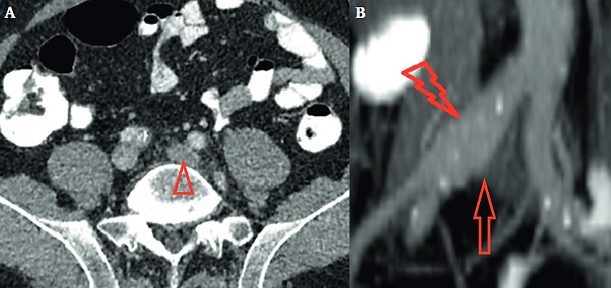
A Rare Case of a Small Iliac Aneurysm Causing Iliac Vein Thrombosis
Spyros Papadoulas et al. Eur J Vasc Endovasc Surg. 2020 Apr;59(4):673 Department of Vascular Surgery, University of Patras Medical School, Patras, Greece
A 66 year old man presented with a two day history of severe left leg oedema. Lower extremity colour duplex ultrasound was negative for deep vein thrombosis. Abdominal computed tomography revealed dilatation of the left iliac venous axis with oedema of the surrounding fat and heterogeneous luminal opacification, findings suggestive of deep vein thrombosis (A, arrowhead). Surprisingly, the cause was compression of the left common iliac vein near its confluence with the inferior vena cava (B, arrow) by a 2.2 cm right common iliac artery aneurysm (B, notched arrowhead). The patient was managed with anticoagulation and elastic stockings.

A Mycotic Saccular Aneurysm Diagnosed With 18F-Labelled Fluoro-2-Deoxyglucose Positron Emission Tomography/Computed Tomography Scanning
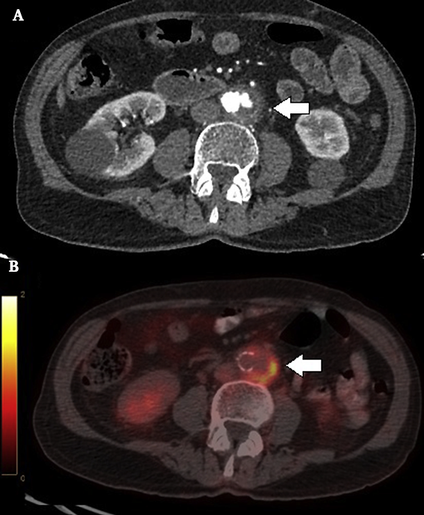
Spyros Papadoulas et al. Eur J Vasc Endovasc Surg. 2019 Nov;58(5):670 Department of Vascular Surgery, University of Patras Medical School, Patras, Greece
An 83 year old man presented with low grade fever, anorexia, and paraumbilical pain. Creactive protein levels and erythrocyte sedimentation rate were elevated, but the white cell count was normal. Abdominal computed tomography (CT) angiography revealed a 3.5 cm saccular aneurysm at the aortic bifurcation (A, arrow). Positron emission tomography with 18F-labelled fluoro-2-deoxyglucose integrated with CT revealed increased metabolic activity in the aneurysm sac. The peri-aortic and prevertebral fat showed higher density, suggesting a mycotic aneurysm (B, arrow). Blood cultures were negative. For family reasons, the patient was transferred to a centre in his home city, where he underwent endovascular repair.

A Stent Graft Visualised Through an Infected Haemodialysis Graft Pseudoaneurysm
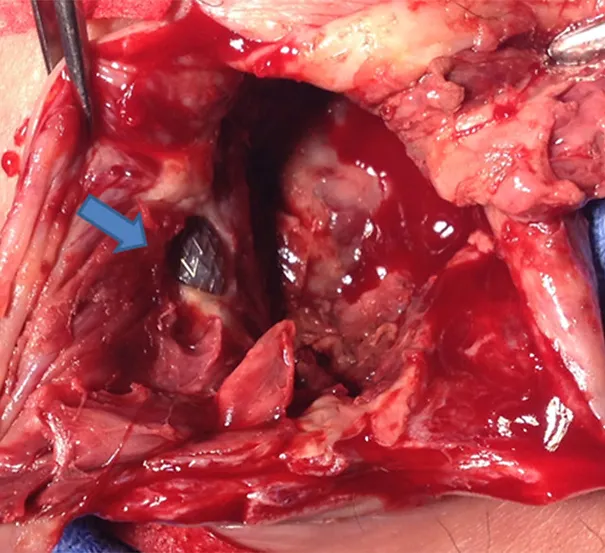
Spyros I. Papadoulas et al. Eur J Vasc Endovasc Surg (2019) 57, 149 Department of Vascular Surgery, University of Patras Medical School, Patras, Greece
A 64 year old male presented with two needlestick pseudoaneurysms of a brachio-axillary PTFE haemodialysis graft; these were repaired by endovascular means using 7 x 40 mm and 7 x 60 mm Covera stent grafts (Bard, Tempe, AZ, USA) with peri-operative teicoplanin antibiotic prophylaxis. Two weeks later he presented with infection of the largest (5 cm) pseudoaneurysm, which was managed by partial graft excision. Intra-operatively the pseudoaneurysm was incised and pus drained. The stent graft was visualised through the graft defect (arrow). The aneurysm sac, part of the graft, and stent graft were excised. Culture results were normal. The wound healed by secondary intention, after a six week vancomycin/ciprofloxacin antibiotic course.

Fever of Unknown Origin due to a Mycotic Abdominal Aortic Aneurysm First Diagnosed with Bone 99mTc Scintigraphy.
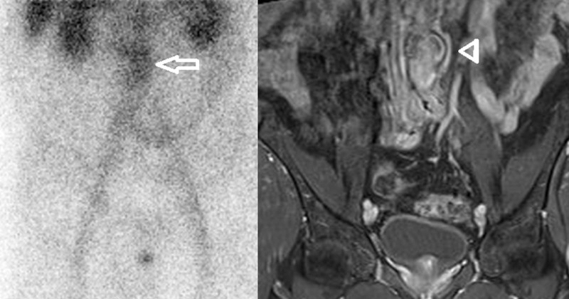
Papadoulas SI, et al. Eur J Vasc Endovasc Surg. 2018 Nov;56(5):698. doi: 10.1016/j.ejvs.2018.08.045. Epub 2018 Sep 29. PMID: 30279059. Department of Vascular Surgery, University of Patras Medical School, Patras, Greece
A 62 year old male, with fever of unknown origin, lumbar pain, weight loss, anaemia, and elevated C-reactive protein/erythrocyte sedimentation rate (CRP/ESR) levels, had bone 99mTc scintigraphy revealing increased 99mTc uptake in the infrarenal aorta (A, arrow). Lumbar magnetic resonance imaging scanning (sagittal short-TI inversion recovery sequence [STIR], T1WI, T2WI, and post intravenous (IV) gadolinium T1WI with fat suppression) showed a 3.8 cm eccentric aneurysm with irregular borders and irregular wall enhancement (B, arrowhead) suggesting a mycotic aneurysm. The aneurysm was repaired with an omentally wrapped PTFE graft. Antibiotics were given for 2 weeks pre-operatively and 6 weeks post-operatively. Tissue cultures were negative. At three month follow up there were no signs of infection.

Listeriosis infection of an abdominal aortic aneurysm in a diabetic patient.
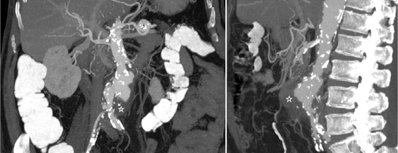
Papadoulas SI, et al. J Glob Infect Dis. 2013 Jan;5(1):31-3. doi: 10.4103/0974-777X.107173. PMID: 23599616; PMCID: PMC3628232. Department of Vascular Surgery, University of Patras Medical School, Patras, Greece
A rare case of an abdominal aortic aneurysm (AAA) infected by Listeria monocytogenes in a 72-year-old male diabetic farmer, is reported. Our patient had a history of a recent pneumonia that could have been caused by Listeria too. Aneurysm infection was manifested by fever and abdominal and back pain, which prompted investigation with CT scanning that revealed a 4.9 cm AAA with typical signs of infection. He underwent urgent AAA repair with aortobifemoral bypass grafting and had an uneventful course. Aneurysm content microbiology revealed Listeria monocytogenes and following a 9-week course of antibiotics our patient remains asymptomatic 11 months later.

Retained subintimal pellet in a carotid artery.
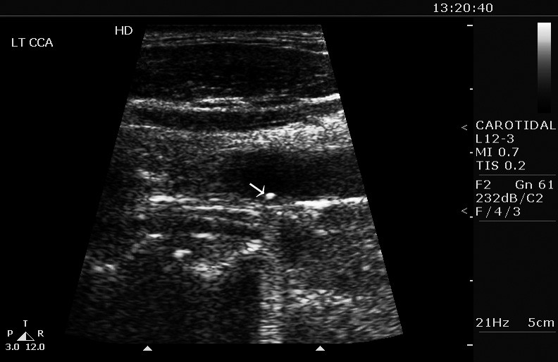
Manousi M, Sarantitis I, Papadoulas S, et al. J Cardiovasc Ultrasound. 2011 Jun;19(2):105-6. doi: 10.4250/jcu.2011.19.2.105. Epub 2011 Jun 30. Erratum in: J Cardiovasc Ultrasound. 2011 Sep;19(3):168. PMID: 21860728; PMCID: PMC3150695. Department of Vascular Surgery, University of Patras Medical School, Patras, Greece
A shotgun pellet is depicted in the present image in a carotid artery under the intima, which remained intact without local complications for up to six months. There is lack of data regarding the natural history of such a carotid pellet, but the experience from the myocardium is that, in the absence of infection, completely embedded missiles are usually asymptomatic, tolerated well and may be left in place.

Mycotic aneurysm of the internal carotid artery presenting with multiple cerebral septic emboli.
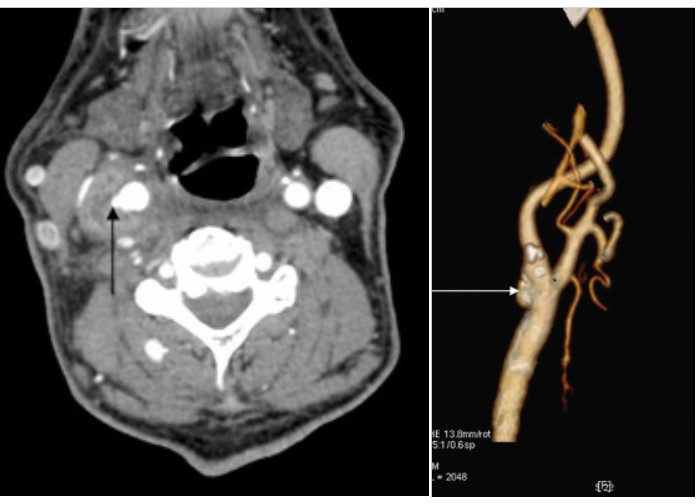
Papadoulas S, et al. Vascular. 2007 Jul-Aug;15(4):215-20. doi: 10.2310/6670.2007.00043. PMID: 17714638. Department of Vascular Surgery, University of Patras Medical School, Patras, Greece
Mycotic aneurysms of the extracranial carotid artery are uncommon and always warrant surgical treatment to prevent eventual rupture and death. Septic embolization to the brain is an even rarer complication of these aneurysms. We present a case of a 79-year-old male with an extracranial internal carotid artery mycotic aneurysm during Staphylococcus aureus bacteremia. He presented with hemiparesis owing to brain embolism from multiple septic emboli originating from the aneurysm. Multidetector computed tomographic angiography contributed to the diagnosis. Resection of the aneurysm and restoration of arterial supply to the brain by vein graft interpositioning was the therapeutic procedure along with long-term antibiotic treatment. A high index of suspicion is required for the diagnosis of a mycotic carotid aneurysm and aggressive treatment is always needed to prevent lethal complications.

Prospective evaluation of biliopancreatic diversion with Roux-en-Y gastric bypass in the super obese.
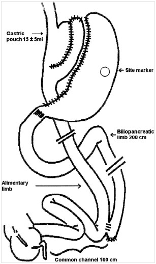
Kalfarentzos F, Papadoulas S, et al. J Gastrointest Surg. 2004 May-Jun;8(4):479-88. doi: 10.1016/j.gassur.2003.11.022. PMID: 15120374. Department of Surgery, University of Patras Medical School, Patras, Greece
The aim of this study was to determine prospectively the efficacy and safety of the biliopancreatic diversion with Roux-en-Y gastric bypass (BPD with RYGBP) procedure used as the primary bariatric procedure in super obese patients. The main characteristics of the BPD with RYGBP procedure were a gastric pouch of 15 +/- 5 ml, biliopancreatic limb of 200 cm, common limb of 100 cm, and alimentary limb of the remainder of the small intestine. From June 1994 through July 2003, 132 super obese patients (body mass index [BMI]: 57 +/- 7), with an incidence of comorbidities 6 +/- 2 per patient, underwent BPD with RYGBP and subsequent follow-up. Mean follow-up time was 29 +/- 14 months. Maximum weight loss was achieved at 18 months postoperative with average excess weight loss (EWL) 65%, average initial weight loss (IWL) 39%, and average BMI 35 kg/m(2). Thereafter, a decline was observed with EWL stabilizing at around 50%, IWL at around 30%, and BMI at around 40 kg/m(2), respectively, by the end of the study period. The majority of preexisting comorbidities were permanently resolved by the 6-month follow-up visit. Early mortality was 1% and early morbidity was 11%. Late morbidity was 27%, half of which was due to incisional hernia. Deficiencies of microelements were mild and successfully treated with additional oral supplementation. The incidence of hypoalbuminemia was 3% and there were no hepatic complications. We conclude that BPD with RYGBP is a safe and effective procedure for the super obese with few metabolic complications.

Vascular injury complicating lumbar disc surgery. A systematic review.
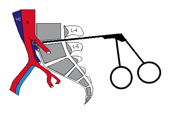
Papadoulas S, et al. Eur J Vasc Endovasc Surg. 2002 Sep;24(3):189-95. doi: 0.1053/ejvs.2002.1682. PMID: 12217278. Department of Vascular Surgery, University of Patras Medical School, Patras, Greece
Objectives: to review the literature concerning the early and late vascular complications of lumbar disc surgery. Methods: using the MEDLINE database, we reviewed all reports of vascular complications associated with surgical excision of a prolapsed disc via a posterior approach reported in the English literature since 1965. Results: we identified 98 cases of vascular complications for an incidence of 1-5 in 10000 disc operations. Early presentation is shock due to rupture of a large retroperitoneal vessel. Late complications include development of pseudoaneurysms and arteriovenous fistulas. Treatment of a vascular tear consisted mainly of primary suturing of the injured vessel. The preferred method for arteriovenous fistula and pseudoaneurysm repair was suturing from within the arterial lumen along with interposition grafting. Recently, endovascular techniques have been recommended, lowering the high morbidity and mortality related to conventional repair. Conclusion: iatrogenic vascular injury during lumbar disc surgery, although rare, should be suspected if signs of circulatory instability are noted or if lumbar pain, leg oedema or high output cardiac failure develop months to years following such surgical procedures. However, these symptoms may arise during or immediately after surgery, requiring immediate intervention.

Ruptured aneurysms of superficial femoral artery.

Papadoulas S, et al. Eur J Vasc Endovasc Surg. 2000 Apr;19(4):430-2. doi: 10.1053/ejvs.1999.0986. PMID: 10801380. Department of Vascular Surgery, University of Patras Medical School, Patras, Greece

Aortocaval fistula in ruptured aneurysms.

Tsolakis JA, Papadoulas S, et al. Eur J Vasc Endovasc Surg. 1999 May;17(5):390-3. doi: 10.1053/ejvs.1998.0777. Erratum in: Eur J Vasc Endovasc Surg 1999 Jun;17(6):554. PMID: 10329521. Department of Vascular Surgery, University of Patras Medical School, Patras, Greece

Objectives: to study incidence, clinical presentation and problems in management of aortocaval fistula in our series.Design:retrospective study.Materials: during a seven-year period, 112 patients operated on for abdominal aortic aneurysm, including four patients with aortocaval fistula.Methods:standard repair of aortocaval fistula from inside the aneurysmal sac was the preferred operative technique.Results:the incidence of aortocaval fistula was 3.6%. Three cases were found incidentally during emergency surgery for ruptured aneurysms; the fourth case was an isolated aortocaval fistula associated with inferior vena cava thrombosis, diagnosed preoperatively by angiography. In this case, inferior vena cava ligation instead of standard aortocaval repair was performed.Conclusions: Aortocaval fistulas, although rare, should be kept in mind, because clinical diagnosis is often difficult. Furthermore, unsuspected problems during repair may necessitate appropriate change in operative technique.